The intervention happens in 3 steps:
Positioning the target-areas
- Positioning the target-areas
- Placing the lead
- Placing the stimulation generator
Positioning the target-areas
An encephalic IRM in stereotaxic-conditions is carried out, and is eventually associated with a cerebral CT scan. A stereotaxic frame is, while under local anesthesia, secured on the skull. That metal piece is made up of a rigid bottom and four vertical props who allow the identification of the target cerebral area, in both great precision and in the three dimensions of space.
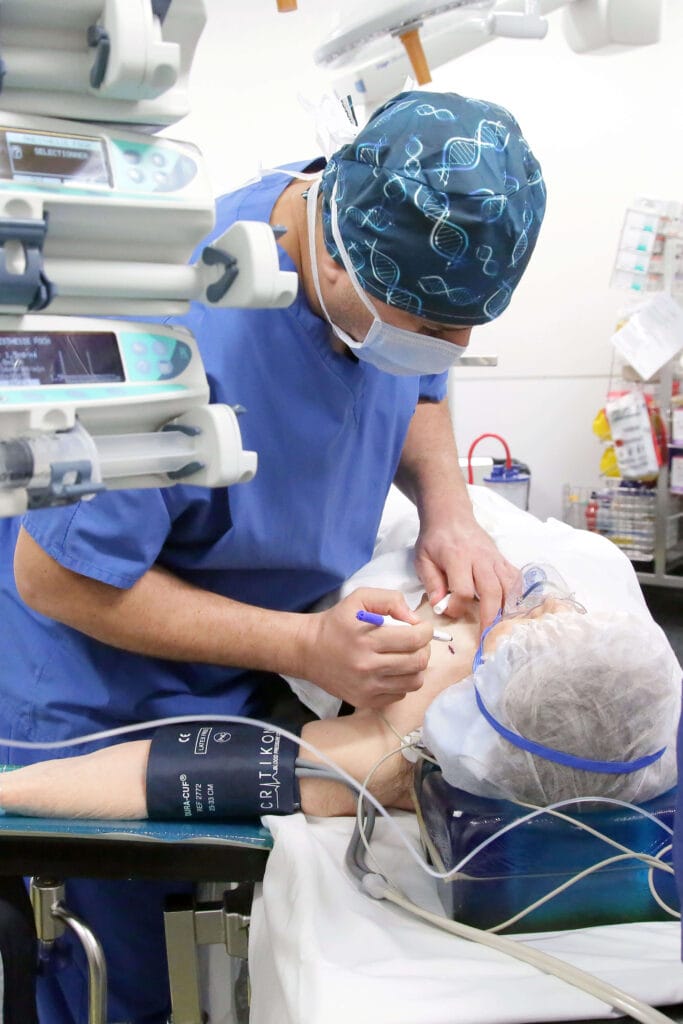
Placing the trial leads
You are then moved once more into the operating room so that the leads can be implanted, under either local or general anesthesia. Trial micro-electrodes can be implanted to record the neurons electrophysiological activity to begin with, and refine the trajectory calculation.
If the patient is under local anesthesia, those micro-electrodes can be used to achieve electric stimulation on the target-area in order to test their impact of the illness’ symptoms. The testing micro-electrodes are then removed and permanent electrodes implanted instead.
Placing the stimulation generator
The electrode is then, either at the same time or a few days later, connected to a neurostimulator which is generally implanted abdominally or in the area under the collarbone, through a subcutaneous connection cable. It is connected to the cerebral electrodes via subcutaneous extensions.